Pages

Monday, January 30, 2012

Delay in the Diagnosis of Rare Diseases

Redirected to: http://whatdnatest.com/delay-diagnosis-rare-diseases/

Rare diseases are usually genetic in origin, life-threatening or chronically debilitating and most of them lack any type of treatment. Also, by definition, a rare disease only affects a small number of individuals, although the exact definition varies from one country to another:
  • In the United States, the Rare Diseases Act of 2002 defines them as "Rare diseases and disorders are those which affect small patient populations, typically populations smaller than 200,000 individuals in the United States." (about 1 in 1,500)
  • While the European Commission definition is "Rare diseases, including those of genetic origin, are life-threatening or chronically debilitating diseases which are of such low prevalence that special combined efforts are needed to address them. As a guide, low prevalence is taken as prevalence of less than 5 per 10,000 in the Community."
Despite the fact that every rare disease only affects a small number of individuals, there are more than 7.000 different rare diseases, so taken together rare diseases affect over 30 million Americans and an estimated 350 million people worldwide according to R.A.R.E. project. However, the diagnosis of rare diseases is not easy and usually there are delays in the diagnosis of these genetic disorders. We can see an example in this video from SWAN UK (Syndromes Without A Name: a project run by Genetic Alliance UK offering support and information to families of children with undiagnosed genetic conditions):

 
(more videos about rare diseases available on the link)

In 2009, EURORDIS published the book The Voice of 12,000 Patients with the results of the surveys EurordisCare2 and EurordisCare3. These surveys were conducted to study the experiences and expectations of patients of rare diseases across Europe regarding access to diagnosis and to health care services. Main findings:
  • 25% of patients had to wait between 5 and 30 years from early symptoms to confirmatory diagnosis of their disease.
  • 40% of patients first received an erroneous diagnosis. This led to medical interventions (including surgery and psychiatric treatments) that were based on a wrong diagnosis.
  • 25% of patients had to travel to a different region to obtain the confirmatory diagnosis, and 2% had to travel to a different country.
  • The genetic nature of the disease was not communicated to the patient or family in 25% of the cases.
  • There was genetic counselling in only 50% of the cases.
  • The average patient required more than nine different medical services over the two-year period preceding the survey.
  • More than one quarter of patients reported difficult, very difficult or impossible access to services. A lack of referral was the most frequently reported cause of impossible access.
  • Moving house and reducing professional activity were some of the daily changes patients and their families were required to make as a result of a rare disease.


In the absence of an accurate diagnosis, questions that the parents need to know usually go unanswered for a long time. Like:
  • What is wrong with my baby?
  • Is the condition going to stabilize or worsen?
  • What can be done to treat the disease or at least alleviate the symptoms?

Many rare diseases show symptoms in the early years, while some others develop at a later age, but in any case they are a matter of concern not only to the patient or the parents but to the whole family. Once a genetic rare disease is detected in a family, other members like aunts, uncles or cousins might want to know if they share this gene and their risk to develop the disease or to transmit it to their children. In order to properly asses this risk, an accurate diagnosis and genetic testing are necessary. However, the doctor that sees the baby (or the adult patient) has probably not seen anyone with this condition before. The patient is refereed to an specialist, but probably he is also unfamiliar with this specific disease. After a number of tests and doctors some families get a diagnosis, some others don't.

A solution to the diagnostic delay would be to increase the awareness, knowledge and coordination of health care professionals on rare diseases, establishing effective referral systems to channel undiagnosed people suspected to be affected by a rare disease to a reference center where he could be diagnosed. Also, more research is needed to better understand each rare disease and enable the development of genetic tests for accurate diagnosis and effective treatments for each condition. To achieve this, it is necessary more awareness from the general public to know about the reality of people affected by these conditions, be more supportive and demand from policy makers the necessary actions. With an estimated incidence of rare diseases of 8-10% as a whole, each one of us is quite likely to have at some point a case of a rare disease among our extended family, friends or coworkers. Let's help them, let's help us.


More information on rare diseases:
  • EURORDIS: the Voice of Rare Diseases Patients in Europe.
  • Genetic Alliance: organization devoted to promoting optimum health care for people suffering from genetic disorders.
  • Orphanet: the Portal for Rare Diseases and Orphan Drugs.
  • NORD: National Organization for Rare Disorders (USA).

More information on rare diseases in Spanish:


This post was published on January 30th 2012, a month in advance of the Rare Disease Day on February 29th 2012, as part of a blog hop organized by R.A.R.E. Project and The Global Genes Project to raise awareness on rare diseases. You may be interested in visiting other participating blogs (at the bottom of this post) and take action:
Global Genes logo for rare disease day on february 29th 2012
  1. Help unite 1 Million for RARE on the Global Genes Project Facebook page so that we can increase awareness to the rare disease community.
  2. Wear That You Care (using jeans to call attention to genes that can cause rare disease) on World Rare Disease Day and encourage others to do so too. Include your schools, sport teams, places of worship, friends, family and coworkers! Share your photos on Facebook. Tag Global Genes Project.
  3. Donate a bracelet to the 7000 Bracelets for Hope campaign and bring hope to a child/family living with rare.
  4. Are you living with rare? Sign up to receive one of the 7000 Bracelets via the Global Genes website and also join the R.A.R.E. network.


Monday, January 23, 2012

Computer Games for Crowdsourcing Scientific Research

Redirected to: http://whatdnatest.com/computer-games-crowdsourcing-scientific-research/

Do you like computer games?

If yes, here you have two options to play and at the same time contribute to genetic and biotechnology research:

example of multiple sequence alignment with regions of similarity highlighted in colors
Figure 1. Example of multiple sequence alignment.
It looks like a game, but it is a tool to improve multiple sequence alignments of DNA regions that may be linked to various genetic disorders. Sequence alignment can be applied to DNA, RNA or amino acids sequences and it is a way of identifying regions of similarity that may be consequence of functional, structural or evolutionary relationship between the sequences (figure 1). This alignment is usually done with the aid of computer algorithms, however they do not guarantee a global optimization as it will take a prohibitively expensive computational power to achieve it.

Humans have evolved efficient pattern-recognition and visual problem-solving skills. Philo abstracts multiple sequence alignment to manipulating color patterns, adapting the problem to benefit from human capabilities. Players receive data which has already being aligned by an algorithm and play to optimize the alignment. With many people working on it eventually some players may end up with a better alignment than the computer. Play philo!

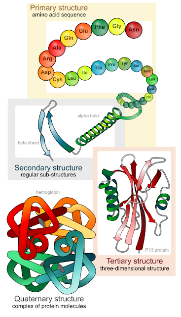
levels of proteing folding: primary structure, amino acid sequence; secondary structure, alpha helix and beta sheet; tertiary structure, three-dimensional structure; quaternary structure, complex of protein molecules
Figure 2. Levels of protein structure.
In this case, instead to multiple sequences alignment, human pattern-recognition and puzzle-solving skills are used to refine protein structures. Proteins allow the cells in your body to do what they do (nutrient transport, metabolic reactions, muscle contractions, chemical signalling, etc). Being able to predict the structure of a protein is key to understand how it works and to target it with drugs in the case that it is involved in a disease.

Proteins are made up of 20 different amino acids and are usually between 100 and 1000 amino acids long. However, they do not look like straight chains of amino acids, they fold up (figure 2) according to the chemical properties of the amino acids and this structure specifies the function of the protein. Foldit allows players to refine the structure of proteins finding more stable configurations. In fact, foldit players have designed a protein that could be useful for the production of renewable fuels, drugs and chemicals with 18-fold higher enzymatic activity than the original.

You can watch the video below for more information on this game. Play foldit!

Friday, January 20, 2012

Mitochondrial Inheritance Pattern

Redirected to: http://whatdnatest.com/genetics/how-genetic-traits-are-inherited/mitochondrial-genetic-inheritance-pattern/

Mitochondrial inheritance is different from the other genetic inheritance patterns in that it has nothing to do with the chromosomes of the father or the mother. Most of the cell DNA is in the nucleus, in the form of chromosomes or chromatin (depending on the level of DNA packing), but a small amount of DNA is inside the mitochondria (figure 1).

The mitochondria (singular: mitochondrion) are organelles inside the cell (figure 2). They carry on the oxidative metabolism of the nutrients and provide the cell with most of the energy that it needs. Due to their specialized function, defects in the mitochondria could produce severe diseases. The origin of a mitochondrial disease could originate from a mutation in the mitochondrial DNA, but could also originate from a mutation in the nuclear DNA since there are very few genes in the mitochondrial DNA and part of the proteins and enzymes of the mitochondria are encoded in nuclear genes, synthesized in the cytoplasm and then imported by the mitochondria. 

When the mitochondrial disease is due to a mutation in a nuclear gene, it will be transmitted according to the other genetic inheritance patterns. However, if the mutation is in the mitochondrial DNA, it will be transmitted with a specific pattern that it is very simple: the mitochondria, and their DNA, are only inherited from the mother. So all the children of a normal (gray color in the figures) father and an affected (orange color in the figures) mother will be affected (figure 3), while none of the children of an affected father and a normal mother will be affected (figure 4).


diagram of mitochondrion: mitochondrial DNA, matrix, cristae, inner and outer membrane, ATP synthase
Figure 1. Structure of a mitochondrion. See the small circular fragments of mitochondrial DNA.

diagram of an animal cell: nucleus, cytoplasm, mitochondrion, golgi apparatus, endoplasmic reticulum
Figure 2. Diagram of a cell. See the nucleus and the mitochondria among other organelles.

diagram of mitochondrial genetic inheritance pattern, father normal, mother has a mutation on mitochodrial DNA, all children affected by the disease, by whatdnatest
Figure 3. Mitochondrial inheritance pattern when the mother is affected.

diagram of mitochondrial genetic inheritance pattern, mother normal, father has a mutation on mitochodrial DNA, all children free of the disease, by whatdnatest
Figure 4. Mitochondrial inheritance pattern when the father is affected.


If you want to read about mitochondrial inheritance pattern in Spanish, you can follow the link herencia mitocondrial.

Friday, January 13, 2012

Structure and Function of the DNA Molecule

fragment of DNA double helix spinning, backbone of sugar and phosphate in green and red, nitrogenated bases in blue
Figure 1. DNA double helix.
Redirected to: http://whatdnatest.com/genetics/structure-and-function-of-the-dna-molecule/

You probably know that the DNA is the physical support of our genetic information and that it is a double helix molecule. Lets see its structure and function with a little bit more of detail.
 
In figure 1 you can see a fragment of DNA. The backbone is in the outside in green (deoxyribose sugar) and red (phosphate) whereas the nitrogenated bases are in the inside in blue. There are four different nitrogenated basis and they always pair up the same way due to their chemical structure: adenine with thymine (figure 2) and guanine with cytosine (figure 3). If we cut a section of the DNA, one nitrogenated base plus one deoxyribose plus one phosphate is called a nucleotide. And a nucleotide and the complementary one in the other strand is called a base pair.
adenine and thymine pairing by hydrogen bonds
Figure 2. Adenine - Thymine.

The pairing of the nitrogenated basis is what holds together the two strands of the DNA double helix. And it is very important for the replication of the DNA: since the pairing of nitrogenated basis is always A-T and C-G, you can separate the two strands of DNA and build a complementary strand for each as there is only one complementary nitrogenated base for each position.
guanine and cytosine pairing by hydrogen bonds
Figure 3. Guanine - Cytosine.

The DNA is a very long molecule and it is highly packed in the nucleus of the cells. In figure 4 we can see how the double helix coils around the histones (the gray balls in the diagram) and then this filament coils and forms the chromatin, that can coil again and again like a rope until it takes the form of a chromosome. However the degree of packing of the DNA varies as the regions that are actively being read or replicated need to be uncoiled.


Functions of the DNA: replication and expression of the genetic information.

  • Replication.
When a cell divides during the growth of the organism, it must pass the same genetic information on to the two daughter cells. As we mentioned above, thanks to the double helix structure of the DNA and the fact that the two strands are complementary, this can be easily achieved. The two strands separate and the enzyme DNA polymerase starts building a complementary strand for each of the parent strands. This way, using each strand as a template to build its complementary the DNA is replicated with a very low error rate (the errors in the replication that are not corrected will cause mutations), and each daughter cell will receive a copy of the genome.

  • Expression.
Thanks to replication and cell division each new cell has a complete set of genetic information, now the important matter is what it does with this information. To grow, develop and carry on with its functions the cell has to read the genes in the DNA and produce proteins and enzymes that are important for the cell's structure and metabolism. This is done in two steps. First the information of the DNA is copied to a fragment of  RNA (a molecule very similar to DNA although it is usually single stranded) by the RNA polimerase, this is called transcription. Then the RNA goes fron the nucleus to the citoplasm and there occurs the second step, the translation of the genetic information by the ribosomes to a sequence of aminoacids that forms a protein. Each gene encodes for a different protein and they are expresed at a different rates and at different times during the life of the cell according to what it is needeed at every moment. In a next post we will explain the expresion and its regulation with more detail.

diagram of DNA packing: double helix, histones, chromatin, chromosome
Figute 4. Packing of the DNA into a chromosome.

Saturday, January 7, 2012

X-Linked Recessive Genetic Inheritance Pattern

Redirected to: http://whatdnatest.com/genetics/how-genetic-traits-are-inherited/x-linked-recessive-genetic-inheritance-pattern/

X-linked recessive is one of the possible ways that genetic traits can be inherited.  This pattern is similar to autosomal recessive genetic inheritance in that one copy of the normal allele is enough to hide the phenotype of the recessive trait. However, since in this case the gene is located in the X chromosome (men only have one X chromosome, while women have two X chromosomes), the gender of the affected parent play a significant role in the inheritance pattern among sons and daughters.

If the altered gene version is responsible for a disease, a man with one altered allele in his only one X chromosome or a woman with two recessive altered alleles will be affected and develop the altered phenotype associated to this disorder (orange color in the figures) instead of the normal one (grey color in the figures). An heterozygous woman with one altered and one normal copies of the gene would not be affected by the disease, but she is a carrier and could pass the altered gene on to her children.

If the mother is affected by a X-linked recessive disease, she will transmit the disorder to all her sons but to none of her daughters (although they will inherit a copy of the altered allele and be carriers of the disease) as can be seen in figure 1:

diagram of X-linked recessive genetic inheritance pattern, mother disease affected, father normal, all sons affected by the disorder, all daughters carriers of the disease
Figure 1. X-linked recessive genetic inheritance pattern when the mother is affected.


However, if the father is the one affected by the X-linked recessive disorder, none of the children will be affected, although the daughters will be carriers (figure 2). When a X-linked recessive carrier woman has children, daughters have a 50% chance of being carriers and sons a 50% chance of being affected by the disease (figure 3).

diagram of X-linked recessive genetic inheritance pattern, father affected by the disorder, mother normal, all sons normal, all daughters carriers of the disease
Figure 2. X-linked recessive genetic inheritance pattern when the father is affected.

diagram of X-linked recessive genetic inheritance pattern, mother carrier of the disease, father normal, 50% sons affected by the disease, 50% daughters carriers of the disorder
Figure 3. X-linked recessive genetic inheritance pattern when the mother is a carrier.


Examples of genetic conditions that are inherited following an X-linked recessive pattern:


If you want to read about X-linked recessive genetic inheritance in Spanish, you can follow the link herencia ligada al cromosoma X recesiva.

Friday, January 6, 2012

X-Linked Dominant Inheritance Pattern

Redirected to: http://whatdnatest.com/genetics/how-genetic-traits-are-inherited/x-linked-dominant-genetic-inheritance-pattern/

X-linked dominant is one of the possible ways that genetic traits can be inherited.  This pattern is similar to autosomal dominant inheritance in that one copy of the altered allele is enough to develop the altered phenotype (orange color in the figures) instead of the normal one (grey color in the figures). However, since in this case the gene is located in the X chromosome (men only have one X chromosome, while women have two X chromosomes), the gender of the affected parent play a significant role in the inheritance pattern among sons and daughters.

If the mother is affected by a X-linked dominant disease, all the children will have a 50% chance of inheriting the disorder, being it equally transmitted to sons and daughters (figure 1). However, if the father is the one affected, he will transmit the disorder to all his daughters but to none of his sons, this is explained in figure 2.

diagram of X-linked dominant genetic inheritance pattern, mother is affected father is normal, 50% children affected, by whatdnatest
Figure 1. X-linked dominant inheritance pattern when the mother is affected.


diagram of X-linked dominant genetic inheritance pattern, father is affected, mother is normal, all daughters affected, all sons normal, by whatdnatest
Figure 2. X-linked inheritance pattern when the father is affected.


If you want to read about the X-linked dominant inheritance pattern in Spanish, you can follow the link herencia ligada al cromosoma X dominante.

Monday, January 2, 2012

The Global Genes Project

I recently came across The Global Genes Project, an interesting rare diseases awareness campaign. Everyone has heard about cancer or aids, but probably not everyone knows about rare diseases. This is because, by definition, a rare disease only affects a few people, some times just some hundreds or few thousands in a whole country. However, there are more than 7.000 different rare diseases, so taken together rare diseases affect over 350 million people worldwide according to R.A.R.E. project.

Rare diseases are usually genetic in origin, life-threatening or chronically debilitating and most of them lack any type of treatment. Please, visit The Global Genes Project to know how can you help to spread the word about rare diseases:

hope it's in our genes, a rare disease initiative

Sunday, January 1, 2012

Autosomal Recessive Inheritance Pattern

Redirected to: http://whatdnatest.com/genetics/how-genetic-traits-are-inherited/autosomal-recessive-genetic-inheritance-pattern/

Autosomal recessive is one of the possible ways that genetic traits can be inherited. In this case, the gene is located in one of the 22 autosomal chromosomes and two copies of the altered allele are needed to develop the altered phenotype (orange color in the figures) instead of the normal one (grey color in the figures). One copy of the normal allele is enough to hide the phenotype of the recessive trait. You can see in figure 1 how both the genotypes AA and Aa yield the same phenotype and all the children of an AA parent will have the normal phenotype.
diagram of autosomal recessive genetic inheritance pattern, one parent affected one normal, by whatdnatest
Figure 1. Autosomal recessive aa x AA

If the altered gene version is responsible for a disease, the affected people have two altered alleles (aa). Heterozygous Aa are not affected by the disease, but they are carriers and could pass the altered gene on to their children. Affected children usually are born to couples where both parents are carriers but not affected by the disease. As can be seen in figure 2, their children have a 25% chance of being affected, 50% chance of being a carrier and 25% chance of not being affected and not being a carrier. It is transmitted equally to sons and daughters and the disease is usually not present in all the generations of the family. If one parent is affected (aa) and the other is a carrier (Aa) the likelihood of having an affected child goes up to 50% (figure 3).

diagram of autosomal recessive genetic inheritance pattern, both parents are carriers, by whatdnatest
Figure 2. Autosomal recessive Aa x Aa.

diagram of autosomal recessive genetic inheritance pattern, one parent affected one carrier, by whatdnatest
Figure 3. Autosomal recessive Aa x aa.

If you want to read about autosomal recessive inheritance pattern in Spanish, you can follow the link herencia autosomica recesiva.